Lecture - 30 General account, ultra structure and nutrition of Bacteria
General account of Bacteria:-


> Bacteria are prokaryotic and unicellular microorganisms.
> They lack nucleus or membrane bound cell organelles.
1. Size:-
- In general, bacteria are between 0.2 and 2 µm - the average size of most bacteria.
- E. coli, a bacillus of about average size is 1.1 to 1.5 µm wide by 2 to 6 µm long.
- Spirochaetes occasionally reach 500 µm in length and the cyanobacterium Oscillatoria is about 7 µm in diameter.
- The bacterium, Epulosiscium fishelsoni , can be seen with the naked eye (600 µm long by 80 µm in diameter).
- Thiomargarita namibiensis (diameter of 750µm) is world’s largest bacteria, a gram-negative.
- Proteobacterium (100—300 µm) found in the ocean sediments off the coast of Namibia.
2. Shape:- Due to the presence of a rigid cell wall, bacteria maintain a definite shape. Shapes may vary in bacteria.
a. Coccus
b. Bacillus
c. Spiral
a. Coccus:- It is round cell.

i. Diplococcus:- The cocci are arranged in pairs. Eg.- Streptococcus pneumoniae, Moraxella
catarrhalis, Neisseria gonorrhoeae, etc.

ii. Tetracoccus:- The cocci are arranged in packets of four cells, as the cells divide in two plains. Eg.- Aerococcus, Pediococcus and Tetragenococcus.

iii. Streptococcus:- The cocci are arranged in chains, as the cells divide in one plane. Eg.- Streptococcus pyogenes, Streptococcus agalactiae

iv. Stapylococcus:- The cocci are arranged in grape-like clusters formed by irregular cell divisions in three plain. Eg.- Staphylococcus aureus

v. Sarcinae:- The cocci are arranged in a cuboidal manner, as the cells are formed by regular cell divisions in three planes. Eg.- Sarcina ventriculi, Sarcina ureae, etc.

b. Bacillus:- It is rod-shaped cell.
i. Monobacillus:- Single bacillus occur alone. Eg.- Bacillus cereus
ii. Diplobacillus:- The bacilli are arranged in pairs. Eg.- Coxiella burnetii, Moraxella bovis, Klebsiella rhinoscleromatis, etc.
iii. Streptobacillus:- The bacilli are arranged in chains, as the cells divide in one plane. Eg.- Streptobacillus moniliformis

iv. Coccobacillus:- These are so short and stumpy that they appear ovoid. They look like coccus and bacillus. Eg.- Haemophilus influenzae, Gardnerella vaginalis, and Chlamydia trachomatis.
v. Palisades:- The bacilli bend at the points of division following the cell divisions, resulting in a palisade arrangement resembling a picket fence and angular patterns that look like chinese letters.
Eg.- Corynebacterium diphtheriae

c. Spiral:- It is spirally curved cell.
i. Vibrio:- They are comma-shaped bacteria with less than one complete turn or twist in the cell. Eg.- Vibrio cholerae

ii. Spirillum:- They have rigid spiral structure. They have typical bacterial flagella. Eg.- Campylobacter jejuni, Helicobacter pylori, Spirillum winogradskyi, etc.

iii. Spirochetes:- They have a helical shape and flexible bodies. Spirochetes move by means of axial
filaments. They lack typical bacterial flagella. Eg.- Leptospira species, Treponema pallidum, Borrelia recurrentis, etc.

d. Others Shapes:-
i. Filamentous:- They are very long thin filament-shaped bacteria. Some of them form
branching filaments resulting in a network of filaments called ‘mycelium’. Eg.- Candidatus Savagella

ii. Star Shaped:- Look like stars. Eg.- Stella humosa

iii. Rectangular:- They are rectangular in shaped. Eg.- Haloarcula spp

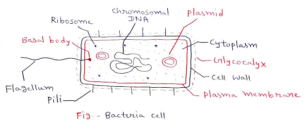
Ultra structure of Bacteria:-

1. Glycocalyx:- This layer is present on the outer surface of cell wall. It is of 2 types -
a. Slime layer
b. Capsule
a. Slime layer:-
> It is an unorganised loosely associated extracellular layer that surrounds the bacterial cell wall.
> It is made up of glycoproteins, glycolipids and exopolysaccharides.
> Slime layers are amorphous in nature and are of varied thickness because they are produced depending on the cell type and environment.
> Because they are loosely associated with the bacterial cell wall, the slime layer can be easily washed off.
> Functions:-
i. It protects the bacterial cell from physical damage such as desiccation and antibiotics.
ii. It helps the bacteria in adhering to smooth surfaces.
iii. A slime layer is mainly composed of polysaccharides and hence is overproduced in unfavourable times as extra food storage for survival.
iv. It is also produced in soil dwelling prokaryotes to prevent them from unnecessary drying during annual temperature and humidity shifts.
v. It sometimes helps the bacteria to survive sterilisation by chemicals such as iodine and chlorine.
b. Capsule:-
> A bacterial capsule is an organised and tightly associated extracellular layer present around the bacterial cell wall.
> It is made up of simple sugars or polysaccharides.
> Unlike the slime layer, it is tightly packed and hence cannot be easily washed off.
> It can be found in both gram positive and gram negative bacteria.
> Function:-
i. The capsules are water loving (hydrophilic) and hence prevent the bacterial cell from water loss or desiccation.
ii. It also protects the bacterial cell wall from engulfment by the white blood cells (phagocytosis).
iii. The presence of a capsule in bacteria determines its virulence factor.
iv. It also helps the bacteria to adhere to various surfaces.
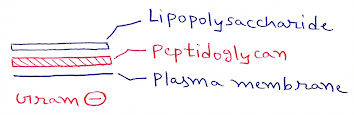
2. Cell wall:-
> It range in thickness around 0.02µ.
> It gives rigidity and shape to the bacterial cell.
> Chemical composition:- The three main constituents of cell wall are:
i. N-acetyl glucosamine (NAG)
ii. N-acetyl muramic acid (NAM)
iii. A peptide chain of four or five amino acids.
- These together form a polymer called peptidoglycan or mucopeptide.
- The NAG and NAM molecules which are arranged alternatively, run in one direction and the peptide chain run crosswise. The rigidity of bacterial cell wall is due to the presence of this polymer.
- Some other chemicals such as teichoic acid, Lipopolysaccharides are also deposited on it.


3. Plasma Membrane:-
> It is about 75 A° thick.
> Chemically it is composed of a double layer of phospholipid molecules.
> Proteins are found embedded in the lipid bilayers.
> Mesosomes:- The membrane has many folded structures called mesosomes which are associated with number of activities like -
i. Site for protein synthesis
ii. Respiratory function
iii. Multiplication of chromosomal DNA
> Plasma membrane contains special receptor molecules that help bacteria detect and respond to
chemicals in their surroundings.
> It also controls the entry of organic and inorganic molecules.
4. Cytoplasm:-
> It is a complex mixture of carbohydrates, proteins, lipids, minerals, nucleic acids and water.
> It stores organic material in the form of glycogen, rolutin and poly-β-hydroxy butyrate.
> The bacterial cell is devoid cell organelles but the photosynthetic bacteria have chromatophores in their cytoplasm.
> Ribosomes are the sites of protein synthesis and suspended freely in cytoplasm. Their number varies from 10,000 to 15,000 in a cell. Bacterial ribosmoes are 70s type (50s and 30s subunits) consists of two subunits.
5. Genetic material:-
a. Chromosomal DNA:-
> The dsDNA molecule is approximately 1,000 µm long, usually forming ring like structure or sometimes remain diffused throughout the cytoplasm of the cell.
> The Bacterial DNA is devoid of histones and referred to as bacterial chromosome.
b. Plasmids:-
> Lederberg (1952) gave the term plasmid.
> These are extra chromosomal ds circular DNA.
> They are self replicative.
> They contain different nonessential characters.
> Based on host properties, the plasmids are classified into different types as:
i. F - plasmid:- F-factor for fertility.
ii. Col - plasmid:- Col-factor for colicinogeny.
iii. R - plasmid:- R-factor for resistance.
iv. Ti - plasmid:- Tumor inducing plasmid (Agrobacterium).
v. Ri - plasmid:- Hairy root inducing plasmid (Agrobacterium).
6. Flagellation:-
> The organ of the locomotion is small whips or hair like appendages called flagella.
> Distribution of flagella:-
i. Atrichous:- Bacteia which lack flagella. Eg.- Lactobacillus

ii. Monotrichous:- One flagella at one end. Eg.- Vibrio chlolerae, Pseudomonas

iii. Amphitrichous:- One flagella at each end. Eg.- Nitosomonas, Spirillum

iv. Cephalotrichous:- Two or more flagella at one end only. Eg.- Pseudomonas fluorescens

v. Lophotrichous:- Tufts of flagella at both the ends, Eg.- Spirillum volutans

vi. Peritrichous:- Flagella distributed evenly all over the body, Eg.- Proteus vulgaris

> Structure of flagella:- The flagella is a helical structure composed of flagellin protein. The flagella structure is divided into three parts:
a. Basal body
b. Hook
c. Filament

a. Basal body:-
- It is attached to the cell membrane and cytoplasmic membrane.
- It consists of rings surrounded by a pair of proteins called MotB. The rings include:
i. L-ring:- Outer ring anchored in the lipopolysaccharide layer and found in gram +ve bacteria.
ii. P-ring:- Anchored in the peptidoglycan layer.
iii. M-S ring:- Anchored in the cytoplasmic membrane
iv. C-ring:- Anchored in the cytoplasm
b. Hook:-
- It is a broader area present at the base of the filament.
- It connects filament to the motor protein in the base.
- The hook length is greater in gram +ve bacteria.
c. Filament:- Thin hair-like structure arising from the hook.
Nutrition of Bacteria:- 2 types of bacteria based upon nutrition -
1. Autotrophic
2. Heterotrophic
1. Autotrophic:- The bacteria which synthesis their own food from the simple inorganic compound, are called autotrophic. 2 types -
a. Chemosynthertic
b. Photosynthertic
a. Chemosynthertic:- These bacteria prepare their food by using chemical energy. They get energy for food synthesis by the oxidation of certain inorganic substances such as ammonia, nitrites, nitrate, ferrous iron, hydrogen sulphides and a number of metalic or non matelic materials avialable in the environment.
i. Sulphur bacteria:- They use chemical energy while there is oxidation of sulphur compound.
Eg.- Thiobacillus
2H2S + O2 ➔ 2S + 2H2O + Energy
ii. Iron bacteria:- They use chemical energy while there is oxidation compound (Fe2+ to Fe3+).
Eg.- Leptothrix, Ferobacillus, Cladothrix
4FeCO3 + O2 + 6H2O ➔ 4Fe(OH)3 + 4CO2 + Energy
iii. Hydrogen bacteria:- They use chemical energy while there is oxidation of molecular hydrogen.
Eg.- Pseudomonas, Hydrogenomonas, Bacillus pectotrophus.
H2 + ½ O2 ➔ H2O + Energy
iv. Nitrifying bacteria:- They use chemical energy while there is oxidation of nitrogen compound.
Eg.- Nitrosomonas, Nitrobacter
2NO2 + O2 ➔ 2NO3 + Energy
b. Photosynthertic:- They can prepare their food by using solar energy in the presence of
photosynthetic pigment bacteriochlorophyll and chlorobium chlorophyll. Photosynthesis in
bacteria differs from other green plants because there is no release of oxygen in photosynthesis.
Such photosynthesis is called anoxygenic photosynthesis. It is of following types:
i. Green sulphur bacteria:- The photosynthetic pigment is chlorobium chlorophyll and sulphur is by-product. Eg.- Chlorobium
6CO2 + 12H2S + Light ➔ C6H12O6 + 12S + 6H2O
ii. Purple sulphur bacteria:- The photosynthetic pigment is bacteriochlorophyll and sulphur is by-product. Eg.- Chromatium
6CO2 + 12H2S + Light ➔ C6H12O6 + 12S + 6H2O
iii. Non-sulphur bacteria:- The photosynthetic pigment bacteriochlorophyll and sulphur is not a by-product. Eg.- Rhodopseudomonas
6CO2 + 12H2 + Light ➔ C6H12O6 + 6H2O
2. Heterotrophic:- They cannot synthesized organic compounds from the simple inorganic substances.
a. Saprophytic
b. Parasitic
c. Symbiotic
a. Saprophytic:-
> They grow in dead, decaying organic material and live by digesting and absorbing them.
> These bacteria gradually break down complex organic compounds into simpler products. For doing so they secreting the enzymes.
> The break down of carbohydrate is called fermentation (Lactic acid bacteria).
> The break down of protein material called putrefaction (Nitrifying bacteria).
b. Parasitic:-
> They live on and within other organisms (host) and they obtain their nutrition from the host.
> They live on or within the organisms both plants and animals.
> If the parasitic bacteria cause diseases and are harmful for their host they are called pathogenic. Many diseases including plant and animal including the man are caused by the pathogenic bacteria. Eg.- Vibrio cholerae, Diplococcus pneumoniae.
> If the parasitic bacteria cannot cause diseases and are harmless for their host they are called nonpathogenic.
c. Symbiotic:-
> They live in close association with other living organisms so that they both are benefited to each other, neither of them is harmed.
> Certain plants establish a symbiotic relationship with bacteria, enabling them to produce nodules that
facilitate the conversion of atmospheric nitrogen to ammonia. Eg.- Rhizobium.
> It appears that not only must the plant have a need for nitrogen fixing bacteria, but they must also be able to synthesize cytokinins which promote the production of root nodules, required for nitrogen fixation.